Looking through you
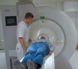
This is how doctors are looking inside your body in the operating theatre. The technique enables doctors to use keyhole surgery for cancer operations that would otherwise require major surgery.
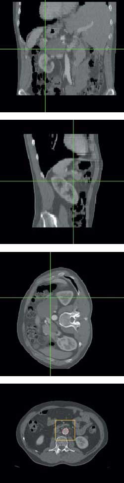
The map images show the way for the operating team during a keyhole operation. The patient has cancer in one of the suprarenal glands and this will be removed. The surgeon can see one of his instruments (white) on the overview graphic together with the surface of the tumour (green), the suprarenal gland (blue) and the main artery (red). At the same time, they can see detailed cross-sections in three orthogonal planes.
Technologists and doctors in Trondheim have collaborated to develop a new IT-based window into the inside of the body. The system converts advanced x-ray pictures and MR pictures to three-dimensional map images that the surgeon can use to navigate when performing keyhole surgery in the abdomen region.
The map images appear on a screen beside the operating table – detailed cross-sections through the body in three orthogonal planes plus general overview images in the form of revolving three-dimensional graphics.
The map images have a common feature: they show where tumours, vital organs and large arteries or veins are in relation to the operating instruments inserted in the patient’s body. In this way, doctors can see far more of the patient’s inside than on video pictures taken inside the body through keyhole surgery. Together with the video pictures, the map images form a valuable extra peephole, according to the pioneers in Trondheim.
“This system will revolutionise parts of
keyhole surgery”, predicts surgeon Ronald Mårvik of St Olav’s Hospital.
In use in the operating theatre
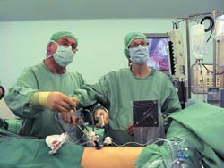
Mårvik, who is one of Europe’s foremost experts on keyhole surgery, has already begun using the newly developed navigation system in the operating theatre.
Mårvik says the system will enable keyhole surgery on tumours that the hospital in Trondheim, like the majority of hospitals, would otherwise have to perform open surgery to remove.
“I am referring to tumours in organs like kidneys, the suprarenal glands and the pancreas and tumours in the thoracic cavities and in the pelvis”, he says. “With this navigation system, we can use keyhole surgery in more such instances than we were able to previously”.
He points out that increased use of keyhole surgery will provide rewards for the individual patient and for society in general. Compared to open surgery, keyhole surgery generates less strain on the patient’s body and, as a result, reduced time in hospital and for convalescence.
Looking «behind» the pictures
When performing a keyhole operation in the abdomen region, the surgeon inserts a thin tube through the skin then in through the abdomal wall. This procedure is carried out from either the front or side of the body. The surgeon controls the operating instruments through the tube, along with a video camera and attached light.
This technique has been used for several years for operations in the stomach and intestine, among other places.
The new navigation system gives the surgeon new possibilities to look behind the video pictures.
Mårvik has already used the system for keyhole operations to remove tumours on the backside of the rear abdomal wall – the part of the abdomal wall that faces the back. The kidneys, suprarenal gland, pancreas and lymph nodes are located here. When the camera is inserted through the abdominal cavity, tumours in these organs are hidden behind the abdomal wall.
“With this navigation system, we are able to see through the rear abdomal wall”, says Mårvik. “In this way, we can see where in the wall we need to cut to find the tumour quickly. We can also see the large arteries or veins on the backside of the wall”.
“Thanks to the navigation technology, we can carry out keyhole operations in this part of the body with an extra high margin of safety”.
SINTEF Unimed, NTNU and St Olav’s Hospital have jointly developed the system. St Olav’s Hospital is among the first in the world to use such a system.
“This solution has attracted the attention of hospitals overseas”, says Mårvik. “Without the rich co-operation between technologists, surgeons and radiologists in Trondheim, we would never have come as far as we now have”.
In a car with satellite navigation, you can trace the car on the map. Through keyhole surgery, surgeons in Trondheim are getting similar help to find their way – inside the patient’s body.
This is how the new navigation system for keyhole surgery works:
The surgeon can follow one of the instruments being used for the operation on map images of the patient’s insides and see where it is located in relation to tumours, arteries, veins and vital organs. Technology from SINTEF Unimed has made this form of navigation in the human body possible. This is how it works in practise:
1. Pictures of the patient
The patient is photographed in an MR (Magnetic Resonance) machine or x-rayed in a CT (Computer Tomographic) machine, in general the day before the operation. These machines take pictures as if the body were divided into many thin slices. Each image shows one of these imaginary cross-sections. A computer program in the navigation system combines the images so that the system has a transparent three-dimensional body accessible. This can be shown to the surgeon in different ways.
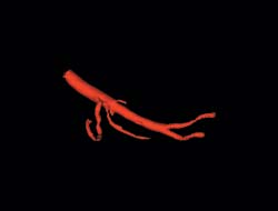
2. Three-dimensional graphics
Each individual image shows a cross-section of the body’s organs. A separate computer program calculates the cross-sections, image by image, and generates a three-dimensional visualisation of the organ. In this case, the program has visualised the patient’s artery.

3. Specially developed pointer
Specially developed metallic pointers are the link between the patient’s body and the graphic image of the body. A sterile version of the pointer is connected to one of the instruments used during the
operation. Three spheres are located on the back of the pointer. Cameras mounted on the ceiling can show at any time where they spheres are in relation to the patient on the operating table.
4. Matching map and terrain
The position of the spheres tells the navigation system the location of the tip of the pointer on the graphic version of the body. The physical world and the image world are matched together before the operation. The patient has marks on their skin from the CT/MR examination and these show up on some of the pictures. Patients receive markers in the same place when they are lying on the operating table. One after the other, the surgeons point increasingly smaller pointers towards these markers.
These pointers also have spheres that the camera can see. From then on, in the image world, the navigation system can recall points that the pointer is directed towards on the patient’s body.
5. Finding the right image
The system selects, from the image library of the patient’s insides, two types of images matching the place the pointer is directed towards:
Three-dimensional graphics that provide overview images. These images enable the surgeon to quickly and effectively see the form and position of organs, tumours and arteries or veins in the patient’s body.
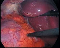
Cross-sections in three orthogonal planes. These two-dimensional pictures provide more detailed information from which the surgeon can navigate. The tip of the pointer appears as a cross in each picture. Here, surgeons Ronald Mårvik and Kristin Helset study images alongside video images from inside the body during keyhole surgery where they will remove a tumour in the patient’s suprarenal gland.
6. Help also before the operation
The map images provide information that can also be used during the planning of the
operation. By using a pointer externally before the operation, the surgeon can see under the skin. This insight can be used to determine where the operating instruments should be inserted. “This is one of the system’s important benefits”, says surgeon Ronald Mårvik of St Olav’s Hospital.
7. Ultrasound next
SINTEF Unimed is now further developing a new navigation system that can be adjusted for use with ultrasound pictures. The picture-based maps that are currently entered into the system show what it looks like in the body before the operation. Ultrasound instruments will update the map images during the operation. “This will, among other things, improve the surgeon’s chances of seeing if they have removed the entire tumour”, explains researcher Thomas Langø of SINTEF Unimed. The institute has previously integrated ultrasound pictures into a navigation system for brain surgery.