Microscopic art and photography: Tora Bardal
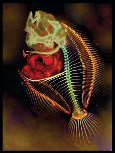
Most fish larvae have nothing but cartilage for their skeletons in the first few weeks of their lives. Fins and calcium skeletons do not develop until after four to six weeks. Many of these larvae are all but invisible to the naked eye. If the larvae grow in poor conditions it may seriously affect their skeletal development.
NTNU scientists are studying what kind of food fish larvae need and how they grow. Skeletal malformations can provide answers to what is missing in fish food or the environment. Bioengineer Tora Bardal photographs thin cross-sections of small organisms. She takes the pictures using a light microscope and magnifying glass. In her spare time, Bardal takes a more artistic approach to the preparations. She does not alter structures or shapes, but uses colours to enhance natural tissue patterns and the microscopic plants and animals themselves.
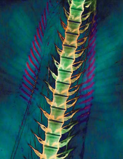
«HERRINGBONE PATTERN» turbot larvae, 25 days
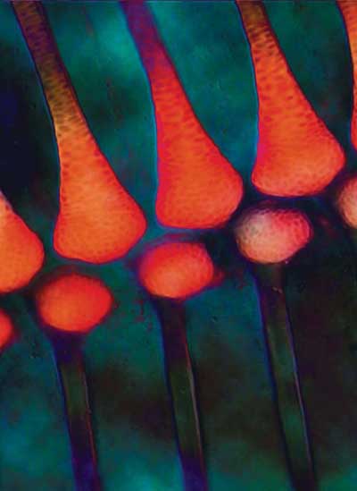
«VERTEBRAE» Cod larvae, 38 days