An eye for detail
The X-ray detectors of the future are on the way. New technology makes it possible to sort plastics, identify useful minerals in waste and reveal contamination in food and medicines.
We have been working on the problem for several years, and we are now getting close to our goal,” says Thor-Erik Hansen of SINTEF ICT.
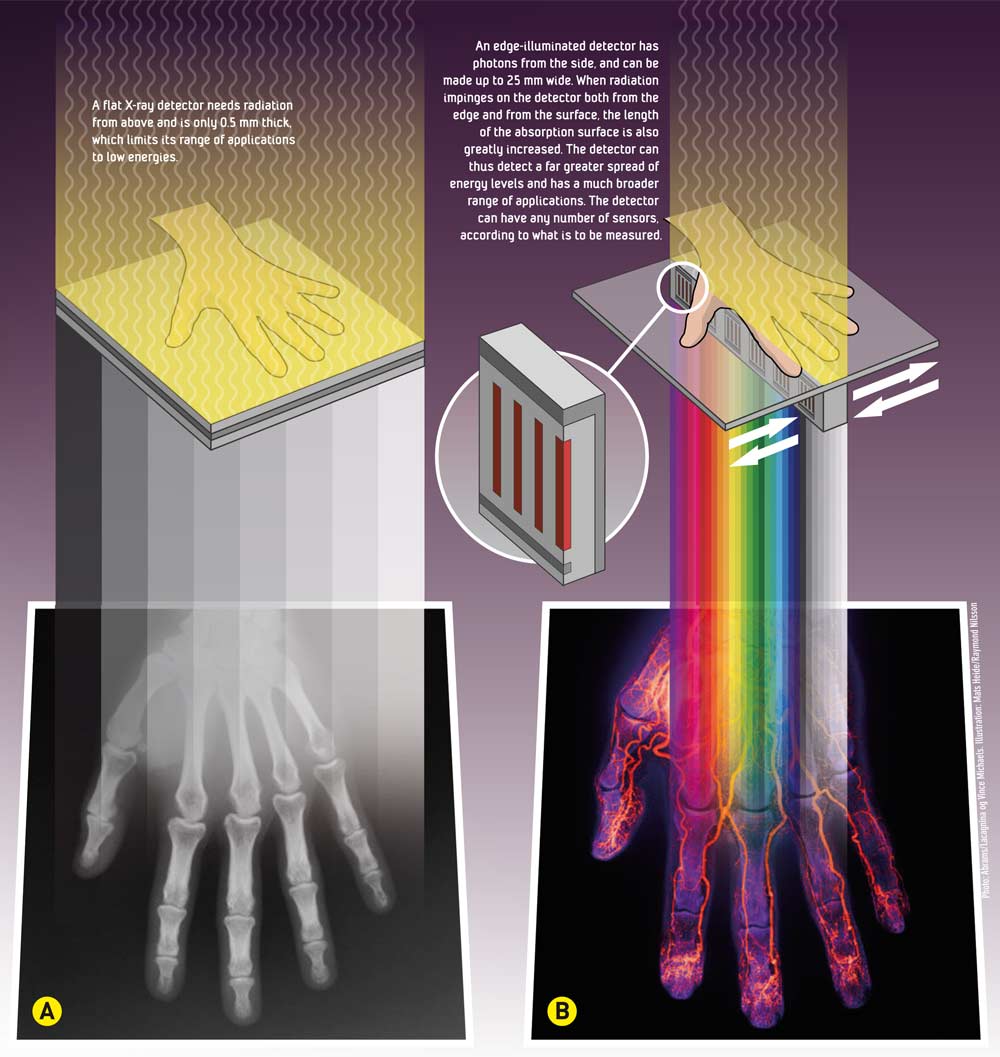
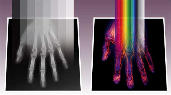
A (left): A flat X-ray detector needs radiation from above and is only 0.5 mm thick, which limits its range of applications to low energies.
B (right): An edge-illuminated detector has photons from the side, and can be made up to 25 mm wide. When radiation impinges on the detector both from the edge and from the surface, the length of the absorption surface is also greatly increased. The detector can thus detect a far greater spread of energy levels and has a much broader range of applications. The detector can have any number of sensors, according to what is to be measured.
For more than 20 years, Hansen and his colleagues at SINTEF’s Micro- and Nanotechnology Laboratory (MiNaLab) have been producing high-energy radiation detectors, which have been used to measure the energy and position of high-energy particles created in physics experiments, such as at CERN in Switzerland. Now, they want to transfer this technology to future X-ray detection systems, employing the same technology there.
The system is based on what are known as ”edge-illuminated detectors”, which allow us to detect a greater range of particle energies than previously.
“In this field, we are world leaders in both development and production,” says Hansen.
The other thing that the detector does is to divide radiation into different energy levels by counting the number of photons and calculating their energy. The new apparatus, which the scientists call EDRD, for Energy-Dispersive Röntgen (X-ray) Detector, also requires lower doses of X-ray energy.
Areas of application
Thanks to their improved contrast, the X-ray detectors of the future will be capable of distinguishing between materials that have very similar densities. For example, they will be able to identify the minerals that would be most profitable to recycle, and to sort soft and hard plastics that could be worth as much as NOK 500 per tonne.
The scientists can also envisage the detector being used as a process-control device in smelters, for estimating the metal content of ores, and detecting hazardous materials in waste or in luggage.
“In the food and pharmaceutical sectors, incorporating x-ray technology in the production line could also reduce the risk of expensive product recalls, since the detector can find contamination or foreign objects in machine-packed products,” says Hansen.
The new X-ray technology can also offer medical benefits. Radiation injury is reduced because of the low doses involved, which can be particularly important in mammography examinations, since these take place relatively frequently.
However, all this will take time. Because the detectors are still at an early stage of development, costs and investments will be involved before the project can reach the application phase.
“In order to address the market and position ourselves as a developer of components, we need first to identify needs and draw up requirements for potential products,” Hansen says.
Today’s x-rays (A)
When you have an X-ray of your hand taken at a hospital today, you lay it on a flat plate. The plate contains the detector itself or, in older machines, the X-ray film. X-ray illumination from an X-ray tube is shone over the hand for a few second to generate the image. The detector pixels under your hand pick up the beam, which is differentially absorbed, depending on the characteristics of the tissue involved.
Our body consists of small molecules (soft tissue) which absorbs little of the radiation, while our skeleton contains calcium – a medium-sized atom that takes up more of the energy of the beam of X-rays. This means that the image of our hand shows the bone structure in white and the soft tissue, which contains carbon, in shades of grey.