Hunting lung tumors
The innumerable divisions of the bronchi often turn the hunt for tumours in the lungs into a game of chance. But soon, lung specialists will be able to navigate accurately inside the airways by “GPS”.
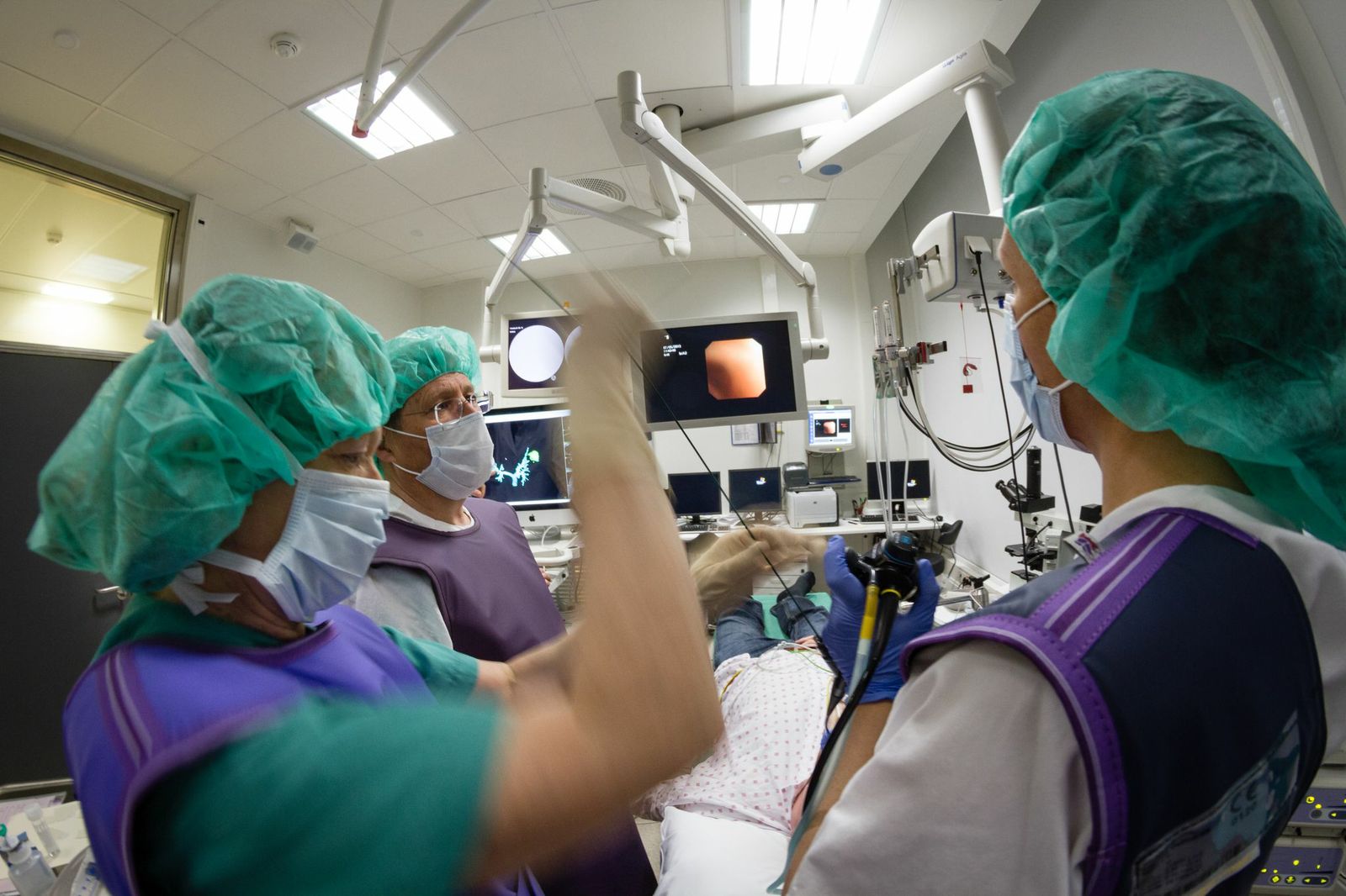
In a bronchoscopy suite at St. Olav’s Hospital in Trondheim, a team of physicians and engineers is in deep discussion with a researcher. The next patient is currently at the focus of their efforts to develop a life-saving technology. He has a tumour in his left lung, and the main challenge is to find out whether it is benign or malignant, precise and quick, at low cost for the patient.
3D navigation
While the patient is prepared with local anaesthetic for the bronchoscopy, the physicians discuss a set of CAT scans that were taken earlier. These are now going to be linked with a completely new diagnostic tool that enables the medical team to locate the tumour with much higher precision than before. The new technique has potential to increase the possibility of a more accurate and rapid diagnosis, and thereby offer the patient appropriate treatment as early as possible.

The challenge lies in getting the system to realize that the pictures that the bronchoscope is taking inside the patient, and the CAT images that were taken earlier, refer to one and the same thing. Photo: Thor Nielsen.
“Lungs are extremely complex organs. We can compare the bronchi to a tree that divides into branches innumerable times. First the trachea itself divides in two in the uppermost part of the chest, then the two main bronchi continue dividing dichotomously – no fewer than 23 times on each side,” explains Håkon Olav Leira, a physician who has recently taken his doctorate degree on image-guided bronchoscopy.This complexity makes it almost impossible to find an easy way through the airways to the site where the medics wish to take their samples. Today, the success rate is between 5 and 30 per cent in cases where the doctors cannot see the tumour, bronchoscopically invisible tumours. Nevertheless, they need to enter the bronchi and take a biopsy.
But now, a navigation system developed by SINTEF researchers and engineers and pulmonologists at St. Olav’s Hospital provides the latter with three-dimensional images of the route into the patient’s lungs and airways, in which the examination will take place. If everything goes according to plan, they will soon be able to localize the tumour within a few millimetres range, with the help of the research scientist Erlend Fagertun Hofstad, the engineer who is co-piloting the navigation at the computer screen.
Camera, map and compass
The tool that the scientists are about to test is known as a bronchoscope, a pencil-thin flexible tube that is fed down the airways and into the bronchi. It is equipped with a camera and a little forceps that enables it to take tissue samples. However, what makes this bronchoscope special is that it is also fitted with a position sensor at the tool-tip, which means that the physicians can always identify the location of the bronchoscope tip in the tissue and bronchi. At the same time, the screen shows them a three-dimensional pre-recorded image of the total “lungscape”, so that they always know exactly where they are.
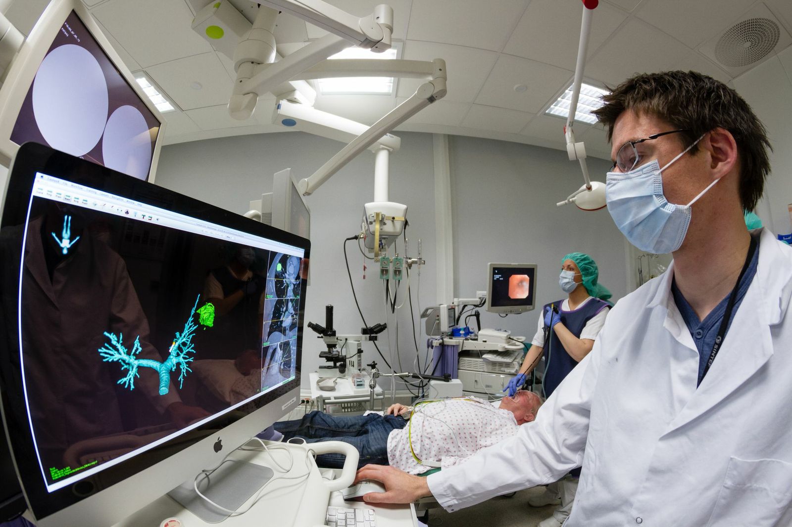
Research scientist Erlend Hofstad checks that “map and terrain” match.
Foto: Thor Nielsen.
“The challenge lies in getting the system to realize that the pictures that the bronchoscope is taking inside the patient, and the CAT images that were taken earlier, refer to one and the same thing. The two sets of images have to fit together like a hand in glove to give us the accuracy that we require. And this has to be done fast. Since the patient is under local anaesthetic but still awake, we have only a certain amount of time to carry out this task,” says Erlend Hofstad.
One of the things that makes this a difficult task is the fact that the lungs are constantly moving through the respiratory cycle; the movements caused by the patient breathing in and out, and those of the bronchoscope itself, mean that the tissue is changing shape and location during the examination. This means that the equipment not only has to take pictures under way but also has to relate these to the CAT images that have already been made. Map and terrain have to match continuously.
Fumbling in the dark
In the examination room the temperature is rising. The patient coughs, and the images on the monitor show that he has started to bleed far down in his windpipe.
“Patients who are taking anticoagulants can be difficult to examine, because they bleed more easily than other patients,” explains Håkon Olav Leira, adding that this demonstrates another of the challenges that the medical team often faces; the bronchoscope images are blurred because blood is obscuring the camera lenses.
“In such situations, it is a huge advantage that we can manoeuvre according to what the position sensor and the 3D map that we have developed in collaboration with the SINTEF researcher tell us. Otherwise, we would just be fumbling in the dark.”
Then the bleeding stops by itself. Once again, everything is under control, and the bronchoscoper soon finds the route through the patient’s bronchi, aided by a steady hand and the images on the screen. It will soon be time to take samples of the 3 cm invisible tumour hidden in the patient’s left lung.
Algorithm
The equipment that is showing the way into the patient’s inner organs also includes a set of advanced mathematical models that have been developed by engineers at SINTEF’s Department of Medical Technology. In practice, the GPS system operates in such a way that the CAT scan is first adapted to the patient, via a registration procedure in which the computer is told that the CAT images of the internal organs must match those of the actual physical patient. The position sensor then registers the movements and position of the sensor that sits at the tip of the bronchoscope.
Facts:
The new navigated bronchoscopy procedure has been developed at SINTEF’s Department of Medical Technology, in collaboration with St. Olav’s Hospital, Departments of Pulmonary Medicine and Medical Technology. The research team has been collaborating closely with the St. Olav’s Hospital doctors for several years and the partnership has resulted in a number of medical innovations in the field of image-guided diagnostics and therapy.
Their solutions offer doctors three-dimensional images of the body’s internal organs while they are operating or performing examinations. For the most part, the techniques utilise ultrasound in combination with CAT images, but magnetic resonance (MR) spectroscopy is another important source of imagery for the three-dimensional “maps”.
The system can then show where the bronchoscope is located on the CAT images. The sensor is less than 1 mm in diameter, and a weak magnetic field that envelopes the patient registers the location of the sensor in three dimensions in real time. Both its direction and position inside the patient are displayed on a screen, so that the doctor always knows where the equipment is.
Successful test
A good hour later, the team has finalized bronchoscopy with different samplings. At her fourth attempt, the bioengineer has managed to get the samples she needs. The team leader is pleased with the efforts of the medics and the scientists:
“That was a good job, well done,” he says to the group at the controls of the equipment, before he thanks the patient for his contribution. Bronchoscopy is a painless, but not exactly pleasant procedure. Willing patients are essential if the scientists are to be able to implement their ideas, and after his efforts, the patient needs some rest.