Secrets of forgetting
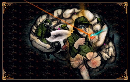
Alzheimer’s disease takes you into a deep darkness. But a laser light and a detective molecule will lead you out.
The mouse will die. It has Alzheimer’s, which means that some protein mol- ecules, slowly and without mercy, will change their structure and become fibrils – useless, thin threads. This process creates irreparable damage. But no one knows what, why and how.
Researchers have given the mouse this illness. But they have also created a tiny window into its small animal brain, through which they can send a special laser light.
The laser sheds light on a scene that to date has rarely been illuminated, and on a life and death drama. Previously, the protein molecule – the bad guy – had the stage all to himself. Now he will soon encounter competition. A young hero is waiting in the wings.
What goes wrong?
The Alzheimer’s puzzle is actually a whole series of riddles. But something links them, this protein that does so much damage on the road to becoming a fibril. What is it that goes wrong?
Proteins are made of small “building blocks” made of amino acid. The cells produce 20 different amino acids, which can be arranged in different ways to determine the kind of protein that we get.
The rows of amino acids that are created inside the cell are like threaded beads on a string that together create a pattern. The pattern is determined by the DNA code. When these strings of beads are combined, they go through a process of folding and forming before creating a protein with its final functional form.
“I believe in 5 to 7 years that we will have come a long way to explaining the mechanisms behind the development of Alzheimer’s.”
Professor Mikael Lindgren
Sometimes this process goes wrong. Misfold- ed proteins are an important part of the prob- lem for many serious diseases: Alzheimer’s, Parkinson’s, type 2 diabetes, Creutzfeldt-Jakob, cancer, cystic fibrosis, and many more.
“We know that something fatal happens along the way. But no one knows in detail why, and how, and how the damage occurs in the process,” says Professor Mikael Lindgren at NTNU’s Department of Physics.
Caught in the act
Researchers at NTNU and the University of Linköping are now at work on the development and use of a so-called molecular probe. This is a specially designed molecule that can be sent into the brain through the blood – a detective molecule, if you will.
Laser spectroscopy, as Professor Lindgren uses it, is all about hunting for the protein misfolding while it is still under way. The detective molecule is the hero in this drama. It changes colour when it finds the criminal. And the spectral frequency of this colour can be observed using a known laser light. Thus the once-darkened stage is brilliantly illuminated for researchers.
Researchers need this kind of help. Once the protein has become a fibril, it is no longer as harmful. The damage seems to occur during the protein’s transition into a fibril. That is when the proteins clump together with other proteins to create a Gordian knot. This brings everything to a stop inside the cells, thus causing the symptoms of the illness. And that is why it is critical to catch the criminal red-handed.
Shuts off vital functions
Yellow illness, red illness...
The detective molecule adheres to a protein tangle. The yellow colour indicates a particular stage of Alzheimer's.
Microscope image: Mikael Lindgren
The detective molecule has a history that extends back to the 1980s. The starting point was something called conducting polymers, which were originally used for batteries, light emitting diodes, and other electronics.
Peter Nilsson, now an assistant professor at Linköping University, discovered several years ago that these polymers can also be used to identify specific protein structures. The polymers are the starting point for the detective molecule now being developed for medical research, in cooperation with various European scientists.
NTNU Professor Lindgren’s specialty is the actual testing of the detective molecule, and creating the colour map that describes what the “detective” has found.
Yellow of a specific wavelength suggests Creutzfelt-Jacobs disease, while another yellow colour may indicate Alzheimer's. Different colours can also indicate a different stage of the disease, because the gradual progression of the illness alters proteins inside the cells. This, in turn is the reason for the different light wavelengths.
The process involves three steps, Lindgren explains: “First, we test these ‘detectives’ – which we also call probes – completely alone with a laser. We learn to get to know them and find out how they react to and use fluorescent light with different wavelengths.
“Then we test how the probes stick to a synthetic variety of an illness molecule, and we develop colour maps based on these results. For Alzheimer’s tangles, the most tightly braided proteins give a yellow light, while a little looser tangle gives more orange-greenish light. The colour phase depends on the type of detective molecule that is used.
“Finally, we send the probe into a living mouse, through an injection into its tail. When we send the laser light through the window in the mouse’s head, we can follow the development of the tangles in the brain. In the future we will be able to do this in a person, but then we will have to use a paramagnetic detective molecule, so that we can follow the process through magnetic resonance imaging,” says Lindgren.
“We know that they eventually turn off some key functions in cells. One possibility is that the cell’s ‘recycling stations’ are damaged or broken. It is these stations that break down unnecessary and damaged molecules and makes them into new things,” explains Professor Lindgren.
“If the detective molecule adheres to the protein on the way to becoming a fibril molecule, and changes colour when it does, we can turn the searchlight onto it and follow it further,” he says.
Seeing the process in colour
The detective molecule is sent into the brain via the blood. It uses the brain’s built-in transportation system, and passes through all of the brain’s defences against intruders. Well inside the brain, it will not relax until it finds a misfolded protein and adheres to it.
When the scientists look with the laser, the detective molecule emits fluorescent light with specific colours. The spectrum can report both that the detective molecule has found its way, and how far the protein thug has come in its destructive process. Different stages of the disease light up in different colours.
“The beauty of the method is that we can follow the illness while it is underway, and learn how the process develops. This is a new approach,” Lindgren adds.
“We are developing a colour image for the disease, so that we can compare it with what develops with medication. We can simply compare the colours of medicated and non-medicated situations over time, and see if different medicines have an effect.”
The professor thinks this approach will provide a much better understanding of the processes in the illness, and could provide the key to finding out how it could be stopped and perhaps reversed.
Needle in a haystack
Many researchers around the world are trying to find the causes of Alzheimer’s disease, and in doing so, hope to find a cure. Most are doctors, while Lindgren is a physics professor who is working with biochemists, among others, to examine the problem from a natural science perspective.
As a physicist, Lindgren has worked with advanced spectroscopy over a number of years. But his interest in Alzheimer’s comes in part from a personal experience.
“My mother suffered from Alzheimer’s and died in 2001. Anyone who has experienced
this disease at close range knows just how much pain and suffering it causes both in the person who has the disease and for everyone around. It was in this context that I decided to approach the disease from my professional point of view,” he explains.
The researchers face a difficult task. There are just a few hundred protein fibrils scattered among a great number of other protein molecules. So looking for them is like looking for a needle in the proverbial haystack. And yet Professor Lindgren is an optimist: “I believe in 5 to 7 years that we will have come a long way to explaining the mechanisms behind the development of Alzheimer’s.”
Can the disease be stopped?
Once this new approach to characterizing Alzheimer’s at the cellular level has succeeded, the trick will be to transfer the knowledge to human samples. But that will require finding an alternative to the window that researchers are able to surgically embed into a mouse brain.
The researchers have considered trying to make a magnetic probe. Thus, the detective molecule could be tracked through the brain by using magnetic resonance imaging. PET scans would also work but are much more expensive than MR.
“With this you can imagine that in the course of 5 to 10 years it will be possible to detect Alzheimer’s at an early stage by using MR,” Professor Lindgren predicts.
The next step will be to create drugs that stop the development of the illness.
Once researchers have been successful in identifying the mechanisms that lead to
Alzheimer’s, it will also be possible to find a drug to combat it.
By Tore Oksholen and Hege J. Tunstad


