New way of visualizing young hearts can save lives
A new kind of ultrasound technology easily shows how blood flows through the heart in foetuses, newborns and children. It’s a tool that can save lives.
The heart is one tough muscle. Even when it is damaged, it continues to beat, as long and as hard it can. Until it can beat no more.
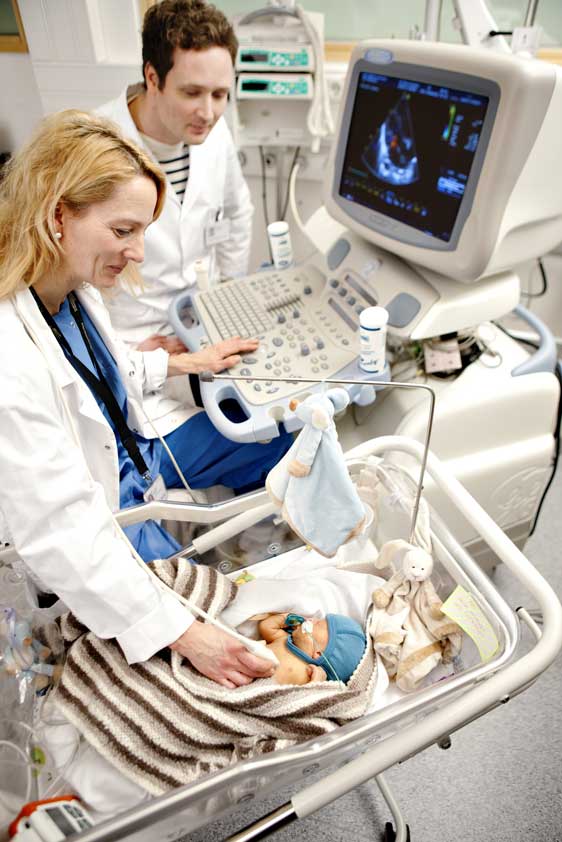
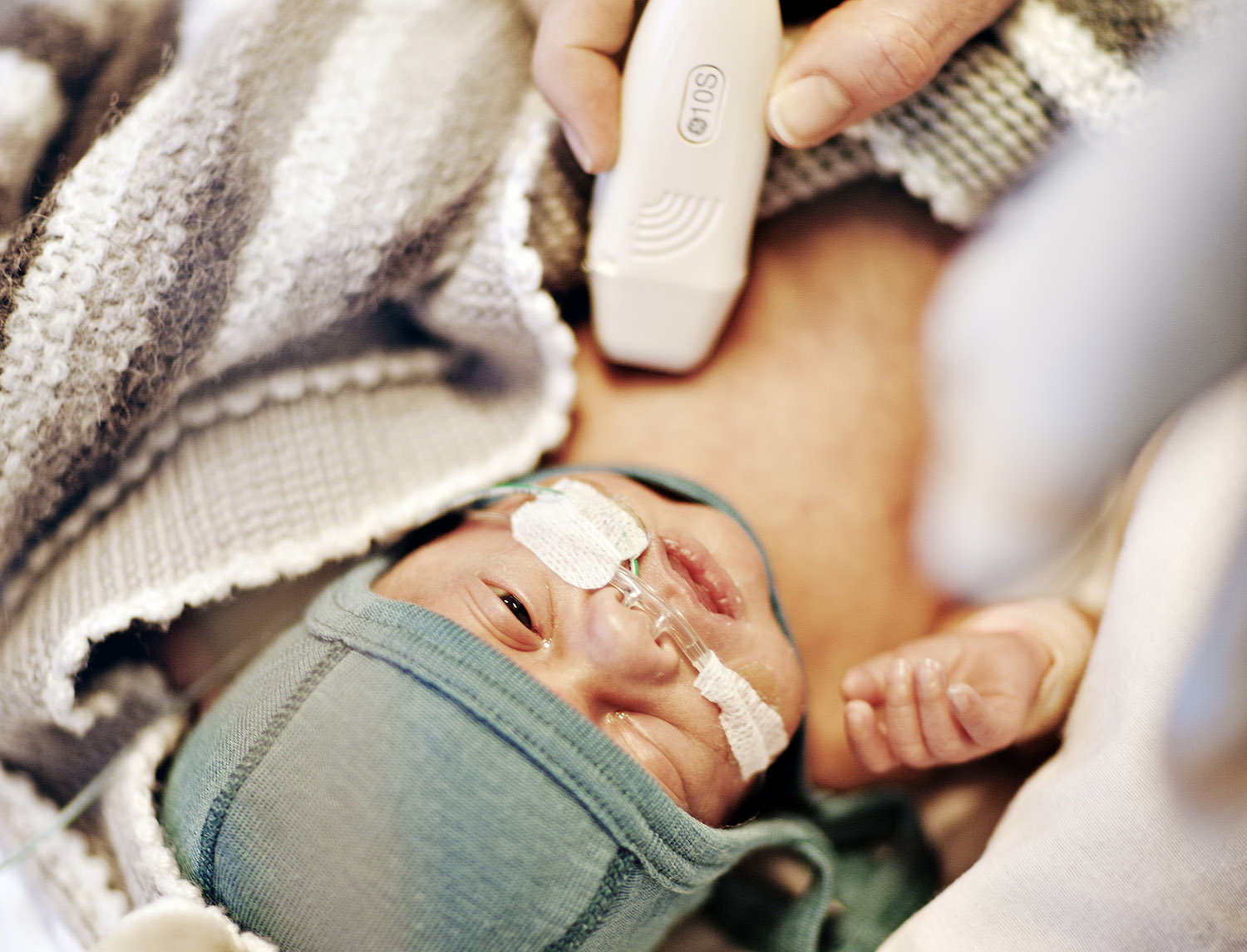
The image on the screen tells Lasse Løvstakken and Siri Ann Nyrnes the condition of this tiny newborn’s heart. Photo: Geir Mogen/NTNU
A damaged heart is constantly trying to compensate for its weaknesses by using its strong and healthy parts to take up the slack for the damaged areas. But this compensation has its costs. Tiny newborns can be blue in the face, even though their hearts are beating. They can be so weak they are unable to nurse.
A modern-day stethoscope
Ultrasound is the doctor’s modern-day stethoscope, providing a non-invasive way to peer into the workings of the heart.
Even with this tool, it can still be difficult to detect problems, and it can be challenging to figure out the reasons for heart failure. For the doctor, the decisive information may involve visualizing how blood flows through the heart – the turns it takes, the kinds of pressures it has along its route, and each of the swirls and eddies it makes.
But until now, no imaging technique has been suitable for examining the detailed blood flow patterns in the hearts of the very young.
Understanding heart disease
“Our hope is to be able to provide ultrasound images that can help us better understand how heart disease develops, which may eventually contribute to a more nuanced clinical diagnosis,” says Lasse Løvstakken, one of NTNU’s Outstanding Academic Fellows and an associate professor in medical imaging at the university.
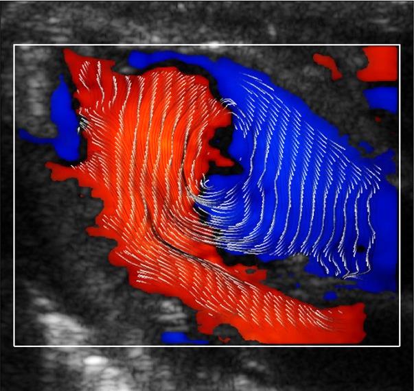
The pattern and direction of blood flow in the heart is very clearly visible with the new technology.
Løvstakken’s new ultrasound technology makes it possible to detect altered blood flow due to heart defects more easily than current techniques allow, by enabling researchers to measure the blood flow in the heart in a more accurate and detailed way. Think of it as a kind of a weather forecast for the heart – at least that is what the ultrasound images actually look like.
Current ultrasound technology uses different colours to depict the blood stream, and the images require interpretation. With Løvstakken’s new technology, the blood’s movement is marked with lines, so that the direction and movement of the blood flow is clearly visible. This has never been done before.
Easy to interpret
The technology has been investigated in newborns. It will now be tested on adults.
Even I, a journalist, managed in less than a minute to see how blood moved through the heart – its direction, as well as all of its small eddies and vortices.
“It is very important that ultrasound images be easy to interpret. The more obvious the imaging is, the better the likelihood of a correct diagnosis. This new technology can be especially important for children, because congenital heart defects are often expressed in altered blood flow,” says Løvstakken.
NTNU's Outstanding Academic Fellows
- NTNU has created a new programme to encourage elite young academics at the university. Seventeen researchers have been selected for the first round of the Outstanding Academic Fellows programme.
- The researchers who have been selected have already made a mark internationally in their respective fields.
- Lasse Løvstakken is one of the 17 fellows.
- The programme provides mentors, meetings, access to research stays abroad, and the expertise of Olympiatoppen, Norway's organization for training elite athletes, to help make them more competitive internationally for grants and research prizes.
Now Løvstakken is working to improve the new technology further, by creating 3D images of how the blood flows through the heart. He will also quantify blood flow characteristics so that doctors can extract numbers that show possible discrepancies. Both of these developments will help doctors to more easily determine if something is wrong, and how large the problem is.
Incorrect diagnoses affect clinical management
Siri Ann Nyrnes, a consultant in paediatric cardiology at St. Olavs Hospital in Trondheim, is collaborating with Løvstakken in a project concerning newborns and children.
“It is important to make the correct diagnosis in a newborn with congenital heart disease. Research has shown that the majority of diagnostic errors in children can affect clinical management. Improved imaging of the blood flow will make it easier make the correct diagnosis, and thereby offer the best treatment and in some cases save lives. However, this connection must be further investigated before we can make any conclusions,” says Nyrnes.