Norwegian superfluid is now helping brain surgeons
The fluid, which resembles brain tissue, makes ultrasound images easier to interpret during an operation. This will make it easier for surgeons to remove brain tumours more accurately.
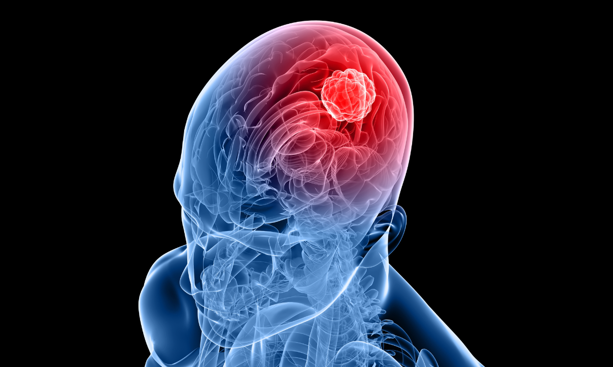
According to the Cancer Registry of Norway, around 1,000 people in Norway are diagnosed with brain tumours every year.
When a tumour needs to be removed, ultrasound is used both to locate it and to see how much of it is left during the operation.
“As a rule, the images are extremely high quality at the start of surgery, but as it goes on, the operation itself creates a lot of noise in the images. This noise makes it more difficult for the surgeon to tell from the ultrasound images how much of the tumour tissue is left and needs to be removed”, explains SINTEF researcher Tormod Selbekk.
Aided by a research team from the National Centre of Expertise for Ultrasound and Image-Guided Treatment, which is a partnership between St. Olav’s Hospital, the Norwegian University of Science and Technology (NTNU) and SINTEF, Selbekk has spent many years trying to find a solution to this problem.
Now he and his team have finally succeeded.
Solving the ‘noise’ problem
The problem is due to the fact that to obtain good ultrasound images, no air must be present between the ultrasound probe and the tissue.
“There needs to be a good connection between the probe and the brain tissue, but as the surgeon gradually removes the tumour, he or she creates cavities that must then be filled with a fluid. The fluid helps to maintain the connection, which makes it possible to obtain an ultrasound image. At the moment, hospitals use a saline solution, but unfortunately this is not the best means of maintaining image quality”, explains Selbekk.
So he and his research team began to look for substances that had the desired acoustic qualities, and that could also be used on human patients without causing harm. Now they have developed a new ultrasound solution using substances that are found in common medicines.
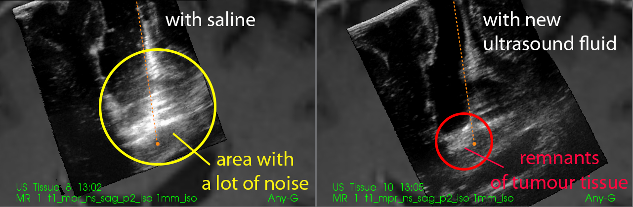
The photos show two different ultrasound images, one of which was obtained using saline solution to fill the operating channel (left), while the other was obtained using the new ultrasound fluid (right). The images were obtained at the end of the operation, when most of the tumour had been removed. At the lower edge of the operating channel, there is a lot of noise in the ultrasound image on the left. In the image on the right, the noise is gone, and you can see a light area at the bottom of the operating channel, which represents the remnants of the brain tumour and must be removed.
Illustration: SINTEF
“We have seen that the new fluid makes it possible to maintain a high image quality throughout an operation. This makes it easier for the surgeons to interpret the ultrasound images and more clearly differentiate between brain tissue and tumour tissue. That then makes it easier to surgically remove as much of the brain tumour as possible, without damaging healthy brain tissue”, relates Selbekk.
According to the SINTEF researcher, this will result in longer survival rates for patients, less damage to healthy tissue, and a better quality of life after the operation.
Works even for the most difficult tumours
“We have always believed that ultrasound is a good instrument for use in brain tumour operations, but a great deal of training was needed to interpret the images when saline solution was used. Noise is a particular problem towards the end of an operation, in the critical phase, which is when the surgeon really needs the best images in order to remove the last remnants of the tumour”, explains Selbekk.
Studies have shown that patients with less aggressive brain tumours have a greater chance of living longer than patients with tumours requiring early and radical removal. The problem with these ‘low-grade’ tumours is that it is difficult to differentiate between tumour tissue and normal brain tissue when looking through a microscope, which is what surgeons in many hospitals do, in the absence of good ultrasound images.
“If you want to remove as much of a low-grade tumour as possible, good quality ultrasound images are an extremely useful tool. Better image quality results in better decision-making than any other method known to us today”, explains professor and neurosurgeon Geirmund Unsgård, one of the researchers working on the project.
No side-effects
The solution has now been rigorously tested in the laboratory. There have been two cycles of animal testing, followed by a clinical study (first phase) on humans, during which three different concentrations of the solution were tested.
“The study showed clear improvements, with virtually noise-free images. The first clinical study also demonstrated no side-effects that could be ascribed to the use of the fluid. Now we need to perform another clinical study, in which we will more thoroughly evaluate the effects and safety of the fluid”, relates Selbekk.
Commercialisation in 2020
Phase two of the study has recently begun. During this phase, the research team will test the fluid using the substance concentration which they believe will achieve the best results. The study will involve a total of 82 patients, and the researchers will use the fluid on one group and the saline on a control group. They hope to have the final results sometime during 2019. The aim is to launch a commercial product in 2020.
“Doctors will be able to use the fluid with every kind of ultrasound machine. This will mean that hospitals all over the world will be able to benefit from the technology, and we believe that this will result in better and safer brain tumour operations in the future”, says Selbekk.
The photos show two different ultrasound images, one of which was obtained using saline solution to fill the operating channel (left), while the other was obtained using the new ultrasound fluid (right). The images were obtained at the end of the operation, when most of the tumour had been removed. At the lower edge of the operating channel, there is a lot of noise in the ultrasound image on the left. In the image on the right, the noise is gone, and you can see a light area at the bottom of the operating channel, which represents the remnants of the brain tumour that must be removed.