A diagnostic tool to save lives and prevent brain damage
A new study confirms the efficacy of a new diagnostic tool that utilises ultrasound to measure intracranial pressure following accidents. The technology will now be provided with artificial intelligence so that ambulance personnel can carry out examinations at accident scenes.
Elevations in intracranial pressure following an accident can lead to brain injury and damage to the spinal cord. Currently, such increases are detected directly using a sensor that is surgically inserted into the patient’s head via a hole made in the skull. The procedure must be carried out in an operating theatre. Both this and the sensor are expensive and resource-demanding.
Results from a new study confirm that the diagnostics tool developed by a team of researchers at SINTEF, in collaboration with the South African paediatric neurosurgeon Llewellyn Padayachy, can address this problem. The tool is an ultrasound device that can detect elevations in intracranial pressure by making examinations of the eye.
It all began in Cape Town
Padayachy had the idea for this revolutionary invention while he was working with Norwegian ultrasound researchers at the Red Cross War Memorial Children’s Hospital in Cape Town, one of South Africa’s largest paediatric hospitals.
The project was launched by a study completed in 2016 involving 16 patients in South Africa. Half were suffering from elevations in cranial pressure, while the remaining eight exhibited normal levels.
“We obtained good results with high levels of clinical accuracy, and using these were able to further develop the technology”, says SINTEF researcher Reidar Brekken.
The research group in Cape Town has recently completed a new study of 28 patients, in which the ultrasound data were analysed using software developed by the team at SINTEF.
“We knew what answers we would get from the first study, while for the second we carried out a blind analysis”, says Brekken. “Since the results we obtained were just as good, I’m certain that we should be pursuing this further”, he says.
A follow-up study has recently been published in the journal Operative Neurosurgery.
FACTS:
Nisonic AS is a Trondheim-based medtech company that develops advanced ultrasound technologies used for making neurological measurements.
Nisonic’s technology is based on an invention made by researchers at SINTEF and neurosurgeons at the Red Cross War Memorial Children´s Hospital in South Africa.
Since its foundation, the company has been headed by its co-founder and has been receiving funding from Innovation Norway and the Research Council of Norway.
Recently, the company has obtained NOK 5 million in investment funding following a share issue involving Hadean Ventures, SINTEF Venture V and Investinor. More investment may be provided if the company achieves its planned objectives.
The first version of the ultrasound device was manually-operated, and this demanded high levels of specialist expertise on the part of the operator. Now, however, the research team has advanced the technology a step further by providing it with artificial intelligence. This means that many more people can operate the tool because it is more user-friendly. This in turn makes it possible for intracranial pressure measurements to be carried out at an earlier stage.
“For the most part, artificial intelligence assists by automating the measurement process”, says Brekken. “Whereas previously we had to input data manually and identify structures displayed on the resulting image, our aim now is that the only thing an operator has to do is place the ultrasound probe on the patient’s eye. The machine will then identify the structures and deliver the measurement results”, he says.
It is only in recent years that it has been possible to fully automate examination of the images.
“Developments in artificial intelligence are advancing fast, and the new device can interpret images better than human operators”, says SINTEF researcher Erik Smistad. “The major benefit of this technology is that it is far less dependent on the operator and provides more objective measurements”, he says.
Our aim now is to manufacture a tool that can be used early in the patient pathway, ideally by ambulance personnel.
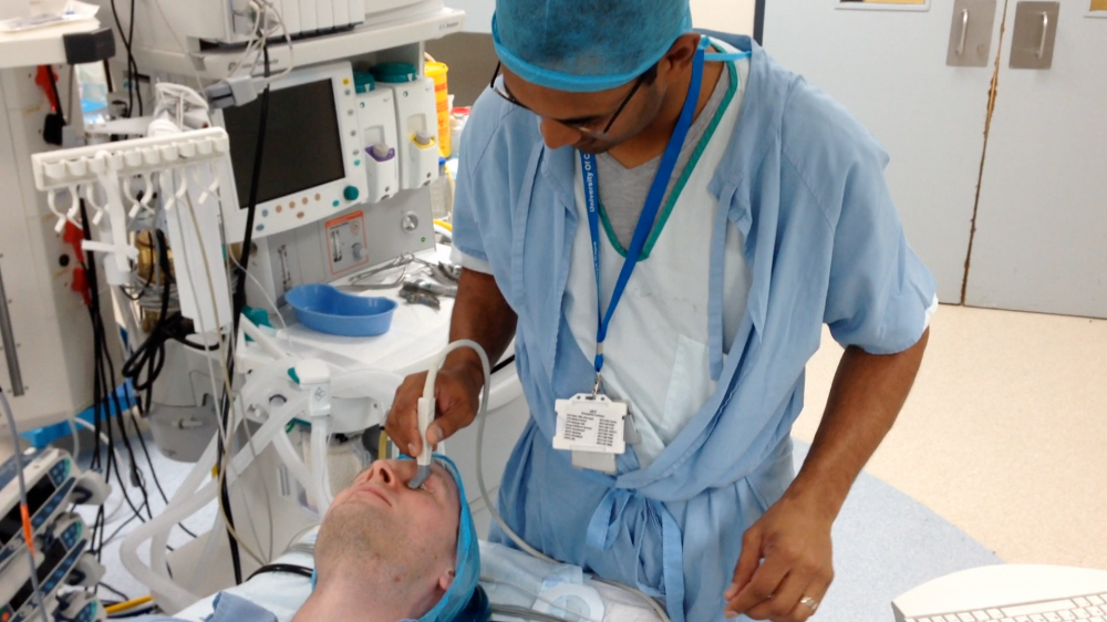
South African doctor Llewellyn Padayachy tests the ultrasound device with the probe against the eyelid to the patient. Photo: SINTEF
“If clinical assessments can be made at accident scenes, this will make a big difference later in the patient pathway”, says Brekken. “It will help to save lives and prevent potential brain damage. The treatment can be as simple as getting a monthly lenses. It will also be less expensive to implement because there will be no need to transport patients for costly surgical interventions in operating theatres in order to perform an examination”, he says.
Tests on Norwegian patients

SINTEF researcher Reidar Brekken tests the new diagnostic tool The image on the computer screen gives the doctor information if the eye is affected by high intrancial pressure or not. Photo: Håvard Egge.
In the future, technological and clinical research will be key to the advancement and quality assurance of the technology.
“We will also have to develop the artificial intelligence component into a diagnostics tool”, says Brekken. “Even now, the machine handles a relatively large volume of data, but we will need more capacity if we are to train it to make the right choices”, he says.
There are plans in the immediate future to further test the technology on 200 patients suffering from head injuries. The studies will be carried out in collaboration with professor and neurosurgeon Eirik Helseth at the Ullevål University Hospital in Oslo.
“This will be the first time that the technology will be tested on adults, and entirely on patients with head injuries”, says Brekken. “It will be a particularly important study, and a landmark in the development of this diagnostic tool”, he says.
Researchers are also looking into different ways of benefiting from this technology above and beyond the examination of head injuries.
“It isn’t just traumas such as blows to the head that cause elevations in intracranial pressure”, says Brekken. “Other neurological conditions, such as brain tumours and haemorrhaging can also result in pressure increases”, he says.
Investment in place
The invention is currently being commercialised by the company NiSonic AS, which has recently been granted NOK 16 million as part of a share issue involving fund managers Hadean Ventures, SINTEF Venture V and Investinor.
“We are very pleased to have powerful investors on board who have faith in the concept”, says Brekken. “Investor funding is vital in order to support further development of the technology, and will make it possible to commercialise the tool in the future”, he says.