New weapon in the fight against liver cancer
A new method can help doctors in planning liver cancer operations.
Liver cancer is one of the most serious forms of cancer. The only possible cure is often to surgically remove the tumour.
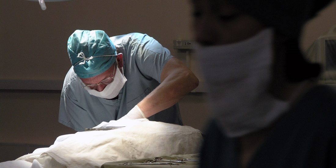
These operations are often carried out by means of laparoscopy, or peephole surgery. In this method, the surgical instruments are inserted into the body in long, narrow tubes, avoiding the need to make larger and more invasive cuts.
Rafael Palomar is a researcher in the Intervention Centre at Oslo University Hospital and in NTNU’s Department of Computer Science. Together with professor and surgeon Bjørn Edwin, who leads the laparoscopic surgery at the Intervention Centre, Palomar has guided the work of creating a three-dimensional model of the liver and tumour, to make it easier for surgeons to find out how to proceed during the resection.
Proceeding as gently as possible is paramount, so as not to damage the liver or the blood supply to it. The liver is vulnerable, and damaging it during an operation can quickly aggravate the situation.
- You may also like: The larger the brain, the greater the risk of a brain tumour
From 2D to 3D
When planning a liver resection, doctors currently have access to two-dimensional images that are always taken. Palomar and his colleagues have developed a computer program that uses the information from these two-dimensional images to create a three-dimensional model instead.
This three-dimensional model can more clearly show the different parts of the liver, as well as the tumour, blood vessels and the surrounding tissues.
“Our method preserves the safety margins better, and the planning time is the same as for other methods,” says Palomar.
The ongoing work has been a collaborative effort that includes Professor Faouzi Alaya Cheikh at NTNU, Professor Edwin at Oslo University Hospital and the Institute of Clinical Medicine at the University of Oslo, consultant Åsmund Avdem Fretland at Oslo University Hospital and Professor Ole Jakob Elle at Oslo University Hospital and the Department of Informatics at the University of Oslo.
Gentler method
The method can help the surgeons use much gentler techniques, because the imaging makes it possible to remove less extra tissue. Surgeons usually remove some extra tissue in the area around a tumour to include any areas where the cancer may have spread but is not yet visible.
Either way, the method is not limited solely to operations on the liver.
“Our results show that the method can be used for all types of surgery and integrated into the planning work,” says Palomar.
Source: A novel method for planning liver resections using deformable Bézier surfaces and distance maps. Rafael Palomar, Faouzi Alaya Cheikh, Bjørn Edwin, Åsmund Avdem Fretland.


