What killed this person?
Sometimes it’s hard to know what a person has actually died from. But post-mortem CT scans may provide a useful tool.
Several researchers at NTNU in Gjøvik are working to figure out how CT images of the dead could be of greatest use.
“We’re trying to identify what factors affect the quality of the CT images,” says David Völgyes, who took his doctorate at NTNU, and who is now a postdoctoral fellow at the University of Oslo.
Occasionally it may be essential for the living to find out why someone died. For example, did a person who was kicked to the ground in town die from a common heart attack? Or were aftereffects from the kick the cause of death? The answer could determine whether anyone will be charged with murder.
Computer tomography, or CT, uses X-ray radiation and is a widely used tool to help figure out various health problems.

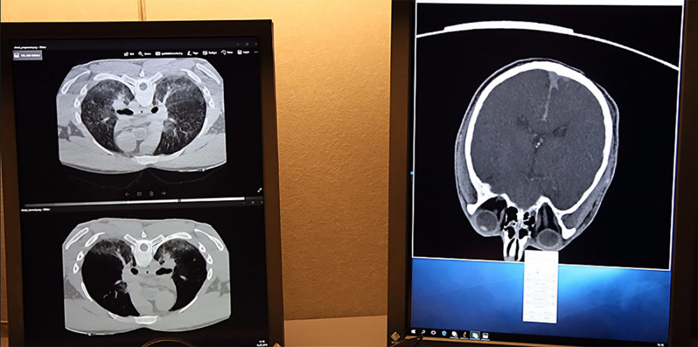
The goal is for pathologists to have another tool to determine cause of death. CT images can supplement traditional exams. Photo: Shutterstock, NTB Scanpix
But CT scans are often not as suitable for identifying different conditions post-mortem. The reason is simply that the body quickly changes after the loss of blood circulation and begins to decompose.
Adding a contrast dye is likewise pointless, because it is transported through the body by the circulating blood in living people. Four articles form the basis for the researchers’ summary of various aspects of CT imaging use.
How to best use post-mortem CT scans
Among other things, the researchers checked more objective criteria for image quality, such as sharpness and contrast. They also had various professionals in the field – three radiographers and three radiography students – evaluate the photos.
“Image quality is highly subjective. It really depends on the preferences of the person who uses them,” says Völgyes.
Sometimes it also depends on what you’re looking for. Searching for dead tissue as a result of a heart attack or from a bullet fragment requires two different approaches.
The research groups used several different procedures and ways of analysing the data to find the best results. They examined the thoracic cavity, but also the head of the deceased individuals.
“It would help if we could use higher doses of radiation post-mortem than we do on living people, but ‘unfortunately’ CT scanners are still designed for the living,” says Dag Waaler, an associate professor in the Department of Health Sciences at NTNU in Gjøvik.
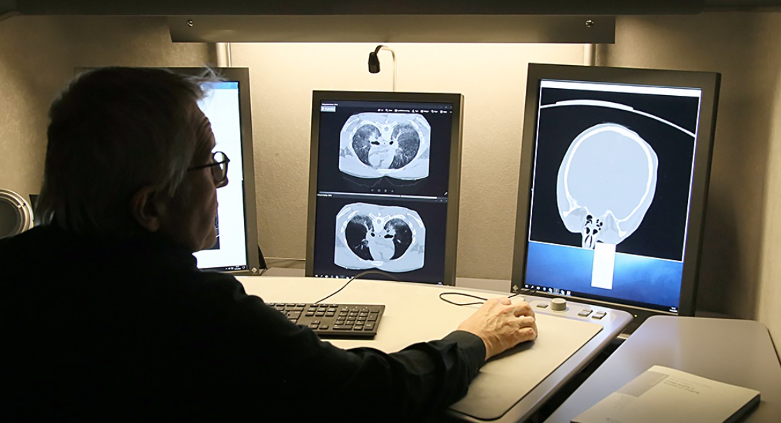
Associate Professor Dag Waaler studies CT images. The quality is subjective, but different factors can still affect it. Photo: Nina Tveter, NTNU
Higher doses can in many cases provide much clearer images. For obvious reasons, you cannot use the same doses for people who are alive.
- You might also like: Children exposed to adult radiation levels from CT scans
New tool
The goal for pathologists is to have another tool to determine the cause of death. CT images have the potential to supplement the use of a scalpel in traditional post-mortem examinations.
This would be welcomed, because things start to change extra quickly inside the deceased once the scalpel is employed.
One challenge is developing clear criteria for how to conduct a CT examination of someone who has died. The ideal result would vary for each individual case.
Nevertheless, the researchers’ conclusion is that CT scans can be useful. The research will also contribute to computers generating even clearer results in the future than are available now.
This research clearly requires teamwork. In addition to Waaler, Völgyes has collaborated with Professor Marius Pedersen at NTNU’s Department of Computer Science, Associate Professor Anne Catrine Trægde Martinsen at UiO’s Department of Physics and Associate Professor Arne Stray-Pedersen at UiO’s Institute of Clinical Medicine, all of whom were also his supervisors.
Source: Image quality in forensic CT imaging. Völgyes, David.