Making ultrasound universally accessible
Currently, ultrasound machines are operated primarily by specialists because it requires extensive experience to interpret the images. Norwegian researchers are aiming to tackle this issue.
The ultimate aim is to develop an ultrasound machine that all health personnel can use without difficulty.
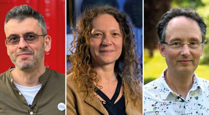
“Such devices must be compact and portable, and thus practical for those working outside the hospital environment – in ambulances, care homes or local doctors’ surgeries”, says Project Manager and Research Scientist Robert Schittny at SINTEF.
Currently, the interpretation of ultrasound images is no easy task. This is why it is only carried out by doctors and others with extensive specialist training and experience in understanding such images.
For this reason, the new device will be equipped with a high level of artificial intelligence that will help users to understand and interpret the images.
A lot of data and limited space
In practice this means that the researchers must gather large volumes of experience-related data from existing ultrasound examinations, and make these accessible in a machine that must also be compact and portable.
The challenge facing the scientists is to select data that is relevant and exclude data that is not, thus ensuring that the data ultimately carried in the device can be processed by a small computer without compromising patient safety.
“Another issue is that it may be necessary to configure the data so that they are adapted to the portable machine”, explains Schittny. “The computing capability installed in a portable device is much less powerful than that in a stationary computer hooked up to hospital equipment”, he says.
It is this aspect of the development process that Norwegian physicists and mathematicians are currently looking into. The equipment will be manufactured by the Norwegian company GE Vingmed Ultrasound.
Helping the user to interpret images
“We will also be providing the device with three-dimensional image interpretation functionality”, says Schittny. “Ultrasound images are of course two-dimensional or “flat”, and this is one of the factors that makes them so difficult to interpret”, he explains.
The system will also be installed with a form of navigation system that will make it easier to understand the anatomy that sources the images we view on the screen.
When we view an ultrasound image of a patient’s abdomen, it is not always easy to distinguish between organs such as the liver or appendix. However, the new device will be equipped to provide the user with aids to understanding such differences. The hope is that this may result in more rapid assistance for patients that need it, and fewer referrals to specialist health services.
“In the future we envisage that a system of this type will enable us to identify areas of concern revealed by the images, such as signs of inflammation or other forms of illness”, says Schittny. “This is the ‘Holy Grail’ that we always carry in the back of our minds, although we don’t know if we will achieve this during this project”, he says.
The research project is called “INHUD”, which stands for INtelligent Handheld Ultrasound Device, and is being funded with NOK 14.2 million over four years by the Research Council of Norway.
The project’s research partners include SINTEF and Norwegian health-tech suppliers GE Vingmed Ultrasound, based in Horten.