The body’s own signalling system provides the key to new diagnostic tools and personalized medicine
Several studies have demonstrated that tiny biological particles called exosomes may carry important information about diseases. We are currently searching for these tiny particles in cooperation with our project partners. If we succeed, we can use the exosomes to predict diseases before they occur.
Exosomes, measuring between about 30 and 150 nm in diameter, occur in blood and other body fluids. Despite their size, almost 1000 times smaller than the diameter of a human hair, they contain a lot of important information about the state of the body. Technologies based on analysis of exosomes may offer hope to those suffering from many diseases, including Alzheimer’s, which is the main focus of the EXIT project.
International research collaboration
As part of a multidisciplinary research project EXIT, SINTEF is collaborating with researchers from several countries to investigate how it is possible to extract exosomes and the information they contain in the lab. This will permit them to be used as disease biomarkers, enabling early diagnosis of a patient’s deteriorating health before irreversible harm is caused. There is currently an ongoing and growing need for medical technology to assist in early personalised diagnoses that will lead to more effective treatments.
How is this possible?
The EXIT project’s working hypothesis is that exosomes, which are associated with neurological function, can be extracted from a patient’s fluid samples using micro- and nanofluidic technologies, combined with a special method for separating and concentrating the particles.
Researchers in the field of micro- and nanofluidics are investigating both the properties and means of manipulation of liquids and gaseous fluids in volumes as small as nano- and pico-litres. The BioMEMS and Medical Sensors Group at SINTEF MiNaLab is currently focusing on the various medical applications of microfluidics that are now possible using the technology developed at MiNaLab.
The method used to separate and concentrate the particles is based on their varying properties and behaviour when subjected to an electric field. To use an analogy, a microfluidic chip acts as a fine-mesh fishing net catching small fish at a specific location in the ocean. The method is based on research carried out at the University of Leiden in the Netherlands, where researchers are developing exosome separation methods from biological samples using nanochannel devices fabricated at SINTEF MiNaLab.
Achieving reproducibility in fabrication of nanochannels
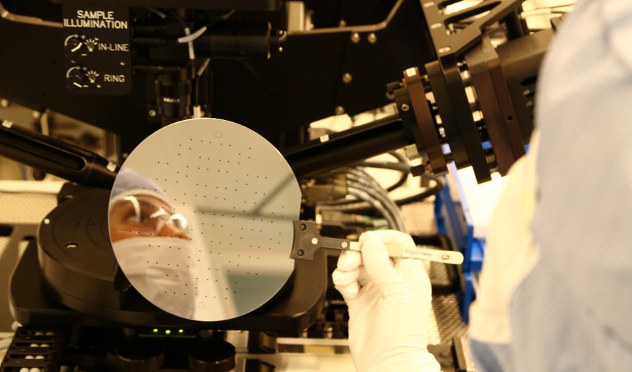
Due to the small dimensions involved, the EXIT project has selected silicon fabrication technology. One of the major drawbacks to the clinical use of micro- and nanofluidic devices has been their lack of reproducibility. Researchers at MiNaLab are now working to develop reproducible fabrication processes for channels as shallow as 40 nm.
The nanofluidic device developed as part of the EXIT project can help to improve current clinical practice by enabling the highly sensitive and high-capacity separation of key diagnostic biomarkers. Robust fabrication processes also promote further advances in micro- and nanofluidics and create opportunities for increased standardization, enabling us to compare results from different laboratories, hospitals and research labs working with exosomes.
If we succeed, the EXIT method may become a very useful tool both in the health sector and in a wide variety of industries. It can be applied in a number of contexts, but already making impact in medical technology and the development of personalized medicines. In the future, we hope that more people will become aware of potential of nanofluidics and nanofabrication.
I would like to thank my colleagues Sigurd Moe, Andreas Vogl and Linda Sønstevold for their excellent input to this blog.


