Artificial intelligence has become the cardiologist’s “super-assistant”
If anything goes wrong with your heart, it is critical to get the right treatment quickly. Artificial intelligence can help with just this – and more lives can be saved.
Cardiovascular diseases are the biggest killers in the world, accounting for 17.9 million deaths globally – every single year, according to the World Health Organisation (WHO). To put that number into perspective, it is more than twice the entire population of London.
So, if there is an urgent need for you to get your heart checked, it is important for the examinations performed by the doctors to be the best they possibly can be.
“One of the most important methods is the echocardiograph,” says Bjørnar Grenne, who is a senior consultant in the cardiac department at St. Olav’s Hospital in Trondheim, and an Associate Professor at NTNU.
Each year, a large number of patients are admitted to St. Olav’s for cardiac check-ups. This means everyone from people with chest pain, to those collapsing in the street, or patients receiving regular heart checks.
What most of them then do is lie on a couch to be examined by a doctor, who uses ultrasound to look inside the body.
“The heart is extremely complex and is very well hidden within the body. We don’t think about it being there, but it’s there all the same, beating up to 100,000 times a day, every day – in each and every one of us.
“There is a good reason why the heart is so well hidden, but because we cannot see it, this also makes it harder to examine. That’s why we need good ways of studying it.”
Needs experience – and a lot of time
The WHO has stated that cardiovascular diseases accounted for 32 per cent of all deaths on a global basis in 2019.
“It’s important to establish what is wrong with the heart at an early stage, so that people can quickly get the right treatment.”
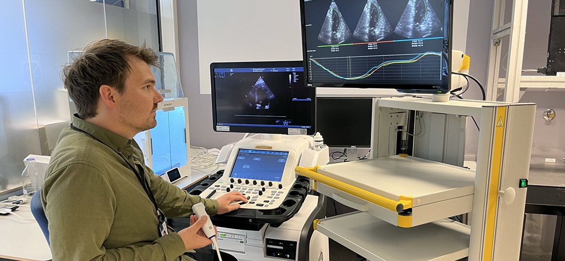
Beside the examination couch, Senior Consultant Grenne demonstrates the probe, which looks like a joystick, and guides it so that the heart of a volunteer from the research team is displayed on the ultrasound screen.
“This gives us real-time images of the heart, which are essential if we are to make the correct diagnosis,” Grenne explains.
The challenge is that you need a lot of experience in order to guide this probe correctly and get the best possible images of the heart. Analysing the images afterwards is also very time-consuming.
“We can take as many as 70–100 different images and videos of the heart during an examination. These must also be studied carefully afterwards by people with a great deal of experience in this field, and that can easily take half an hour if you want to do it properly.”
AI is the doctor’s super-assistant
That is where AI, or artificial intelligence, comes in as an excellent assistant.
“Artificial intelligence can help Bjørnar and his colleagues guide the probe in the right direction and obtain the perfect image every time. AI can also analyse the images as soon as they pop up on the screen and help us to see what is wrong with the heart,” says Andreas Østvik, who is a researcher at SINTEF Digital and NTNU.
Using machine learning, the researchers have fed info into a machine, with Senior Consultant Grenne and his colleagues defining the criteria that must be met in order to get the right cardiac images, and how these images should be interpreted.
This allows artificial intelligence to be used to harvest experience from previous patients, which means that the doctors can make their decisions based on potentially thousands of similar examinations.
During the process, the AI assistant shows a green or red light, so that the doctor knows whether the probe is at the right angle. When the images are correct, AI interprets these, and automatically takes measurements of the heart. Typically, these are measurements of the size of the heart, and how good it is at contracting.
Testing on patients
The technology has now been developed to an advanced stage and has already been used on some patients as part of the treatment offered to them in connection with the research project. However, strict rules apply in the field of medicine, which means that it could take some time before doctors will be able to use this treatment option in all hospitals in Norway.
“We’re expecting it to be available in a few years. We have to test it first – patient safety is paramount, and we have to know that it works before it is made available everywhere,” the SINTEF researcher says.
Grenne adds that the current way of examining the heart is very good, but that it will obviously be tremendously helpful if AI could contribute as an assistant.
“It saves us a lot of time and resources, which means that we can help even more people – which could then save more lives,” he says.
Listen and learn more about the doctors’ use of artificial intelligence on the SINTEF podcast ‘Smart forklart‘.