Ultrasound can prevent brain damage in sick newborns and premature babies
Ultrasound technology from NTNU makes it possible to monitor cerebral blood flow in newborn babies, helping prevent brain damage in premature and sick infants who require surgery.
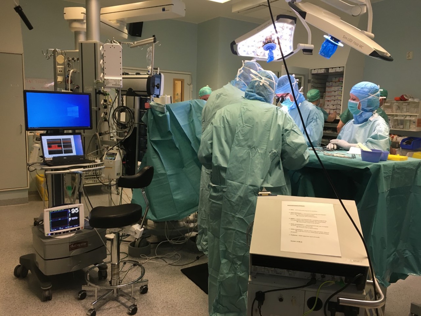
Researchers and neonatal specialists at NTNU and St. Olavs Hospital are behind the first study in which ultrasound is used to monitor cerebral blood flow in newborn babies under general anaesthesia.
“We have always believed that this could revolutionise neonatal care. Everything indicates that we are closer to being able to prevent brain damage in premature infants and sick newborns,” says paediatrician Sigrid Dannheim Vik, who is doing a PhD on NeoDoppler technology.
30 newborn babies monitored during surgery
Sigrid Dannheim Vik. Photo: Siri Ann Nyrnes
Over a two-year period, she has used the equipment to measure cerebral blood flow in 30 newborn babies under general anaesthesia. Some of them were born very prematurely. Others came into the world around their due date, but were born with congenital defects that had developed in the womb.
Most of the patients had gastrointestinal complications, such as lack of passage through the small intestine. Premature babies often have an immature intestine that is easily perforated in the neonatal period.
“These babies require surgery – often acutely. There is no other choice,” says Vik.
The longest courses of continuous ultrasound measurements lasted 10-11 hours, and some of her patients weighed less than 1000 grams.
Very susceptible to brain damage
Regardless of whether they were born very prematurely or have congenital defects and injuries, these are the health service’s smallest and most vulnerable patients.
Babies who are born prematurely are prone to brain damage in the neonatal period and are very vulnerable to blood pressure fluctuations. The risk increases if they have to undergo surgery.
The aim of the NTNU study has been to use the new ultrasound technology to see how cerebral blood flow changes while the infants are under general anaesthesia.
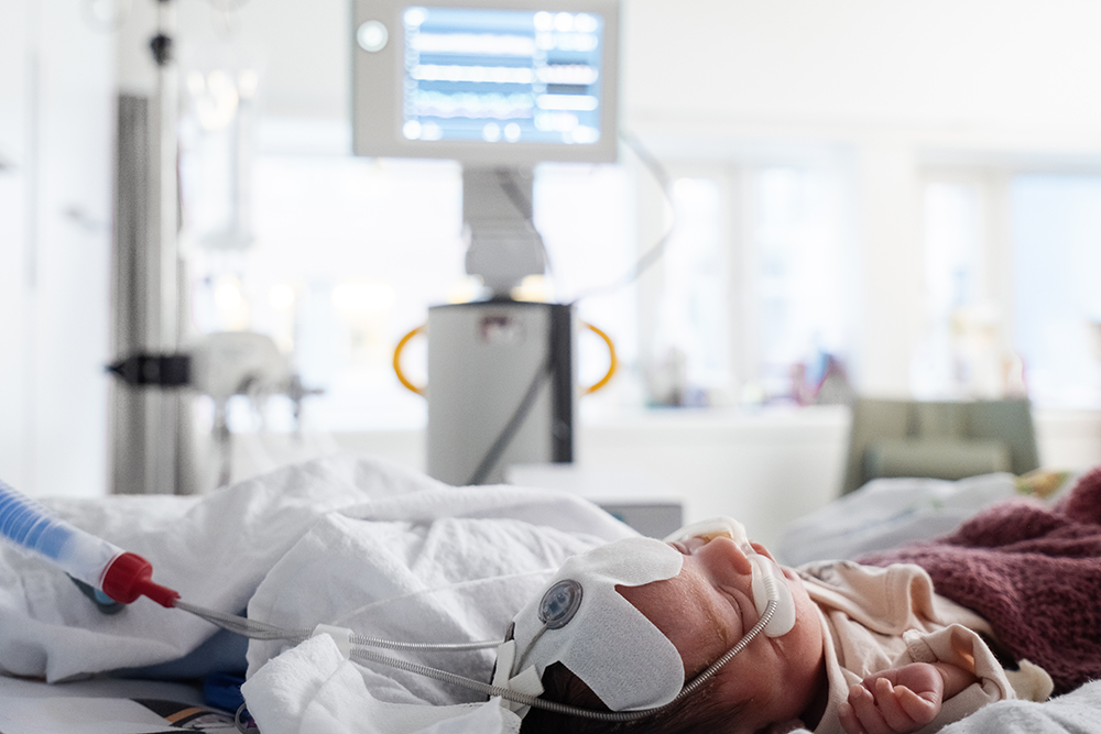
New ultrasound technology can help prevent brain damage in prematurely born and sick infants who need surgery. Photo: Ole A. Ekker
Providing access to completely new information
The conclusion is that NeoDoppler provides doctors with access to critical information that has previously been unavailable.
“The equipment provides important additional information compared with today’s standard monitoring equipment,” Vik said.
Currently, indirect factors such as blood pressure, pulse measurements and general clinical assessments are used to see if the child has adequate blood circulation.
“We don’t actually have a reliable measurement method for monitoring the most important organ, namely the brain,” says the paediatrician and researcher.
This is NeoDoppler - and the people behind it:
- Developed through a collaboration between the Department of Circulation and Medical Imaging (ISB) at NTNU and the Neonatal Intensive Care Unit at St. Olav's Hospital.
- The inventor of the technology is Professor Hans Torp, who is a key figure in the world-leading research community in medical ultrasound in Trondheim.
- Pediatric cardiologist and researcher Siri Ann Nyrnes is responsible for the clinical development of the equipment and serves as the main supervisor in the project.
- Pediatrician Sigrid Dannheim Vik is pursuing an innovation Ph.D., where she conducted the necessary clinical trials before the equipment can be commercially available.
- The technology and equipment were developed with project funding from NTNU Discovery and the Research Council of Norway's FORNY program, totaling around 8 million Norwegian kroner.
- Simultaneously with the trials and clinical studies, efforts have been made for commercialization. NeoDoppler was further developed through NTNU Technology Transfer and is now being commercialized through the company Cimon Medical AS.
Allows rapid implementation of life-saving measures
For anaesthetists, it is all about keeping the blood circulation stable. NeoDoppler measurements are taken continuously before, during and after surgery. The equipment is able to instantly detect changes in cerebral blood flow. As a result, measures that can prevent brain damage and that are adapted to the individual child can be implemented immediately.
It also helps doctors make individual assessments and tailor treatment, rather than having to rely on general recommendations.
“I am in no doubt that we are now moving closer to preventing brain damage.”
May result in new standards
The researchers believe NeoDoppler can provide a new standard in monitoring newborn babies.
“The problem today is that we don’t know what the most favourable blood pressure in newborns is with regard to maintaining stable blood circulation in the brain, and it probably varies from one infant to the next,” says Sigrid Dannheim Vik.
What she and her colleagues saw, and which they believe gives cause for concern, was that cerebral blood flow speed was more than halved during anaesthesia compared with when the patients were awake.
For children who come out of the mother’s womb before week 28 of pregnancy, the chance of brain damage is as high as 50 percent. At St. Olav’s Hospital in Trondheim alone, around 300 children are put under anaesthesia each year before they turn one. Photo: Sigrid Dannheim Vik
“Our findings indicate that newborns who are placed under general anaesthesia should probably have a higher blood pressure than is currently recommended. The current reference range should probably be higher,” says Vik.
A window into the brain
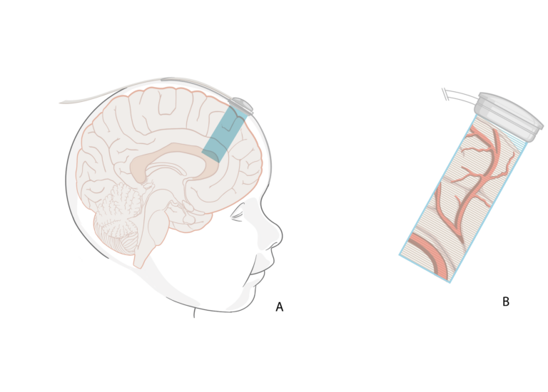
The probe measures the speed of blood flow in large and small blood vessels. The ultrasound waves are sent 3-4 centimeters down into the brain and come back as images that the doctors can see and interpret on a screen. Illustration: Inventas
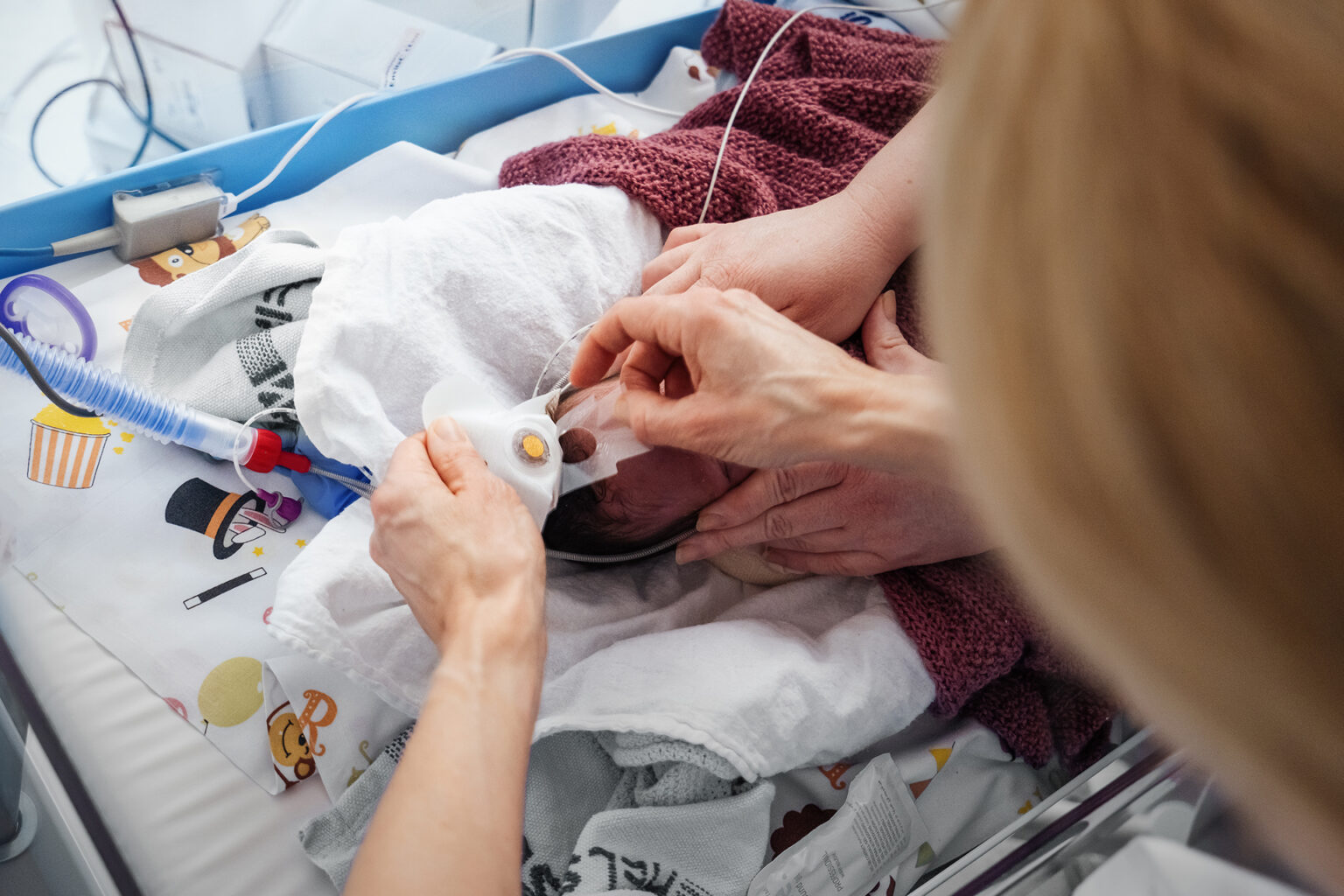
The NeoDoppler ultrasound probe is the shape of a small button and has a diameter of around 1 centimetre. It can be attached to a cap and placed gently over the fontanelle (the soft spot at the top of a baby’s head).
The fontanelle closes towards the end of the first year of life when the bones of the skull fuse together. Ultrasound is not able to pass through bone. Therefore, the fontanelle is like a window that is only open for a limited period of time, through which doctors can directly monitor blood flow.
From mild ADHD to severe movement disorders
Adults have a protective mechanism allowing them to maintain stable cerebral circulation even though blood pressure may vary. Infants do not have this protective mechanism, making them particularly vulnerable to variations in blood pressure, which in turn can lead to unstable cerebral blood flow.
This can also result in the brain being starved of oxygen, which may cause brain tissue damage.
Sigrid Dannheim Vik explains that this can lead to a wide range of brain injuries or delays in development that become noticeable at different ages.
“It can include everything from ADHD and minor developmental disorders to more extensive cognitive and motor difficulties.”
The smallest are most at risk
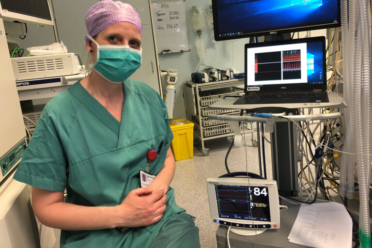
Ultrasonography experts, pediatricians, and neonatologists at NTNU’s Department of Medical Imaging and St. Olav’s Hospital are the first to measure cerebral circulation during anesthesia in premature and critically ill newborns. Photo: Ole A. Ekker.
Every tenth child in the world is born prematurely, which in itself increases the chance of brain damage. For babies born before week 28 of pregnancy, the chance of brain damage is as high as 50 percent.
At St. Olavs Hospital alone, around 300 children under the age of one are placed under general anaesthesia each year.
In addition, there are many other children who are born with congenital defects. The researchers believe that children born with congenital heart defects are another patient group that could benefit from NeoDoppler technology.
Large and varied patient group
In addition, there are children who require surgery due to complications in the neonatal period. For example, there may be perforations in the intestines of premature babies and infants who need anaesthesia due to less invasive surgery or diagnosis.
“In other words, there is a fairly large and varied patient group that can benefit from this type of monitoring,” says Sigrid Dannheim Vik.
An extremely important tool
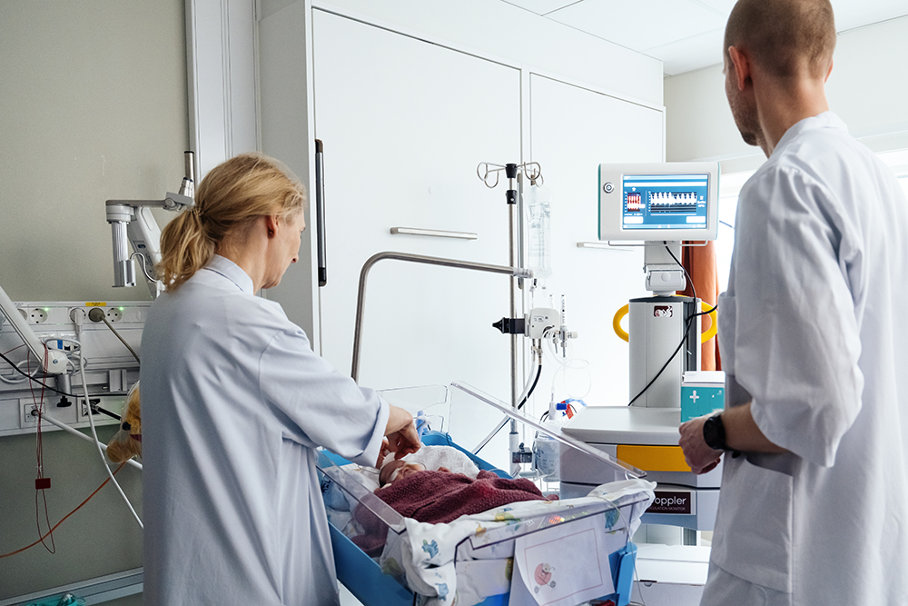
The equipment is lightweight, easy to assemble and use, even for non-experts. It can remain in place for several days and be used wherever the child is at any given time. Photo: Ole A. Ekker.
The paediatrician and doctoral research fellow calls NeoDoppler “a huge advance in neonatal medicine.” She compares it to getting information via colour TV versus listening to the radio.
Where doctors previously relied on indirect measurements, they now have a direct picture of cerebral blood flow.
“It is an extremely important tool – both for the anaesthetist monitoring the child under general anaesthesia and for the rest of us involved in neonatal medicine. It will help us make direct assessments and take measures, thereby avoiding both undertreatment and overtreatment.”
Measuring speed, not quantity
One possible criticism of NeoDoppler is that it measures speeds, resulting in constantly changing images and graphs. It does not measure the exact amount of blood passing through the blood vessels. If the blood vessels vary in diameter, the speeds will also vary.
However, the researchers refer to studies showing that there is a good correlation between speed measurements and the blood flow itself – as long as the speeds are measured continuously.
More long-term studies required
More clinical trials are needed before NeoDoppler becomes standard monitoring equipment in neonatal care. These will be accurate with regard to effect, but the children must be followed up over a longer period of time in order to show that it helps prevent brain damage.
Paediatric cardiologist and researcher Siri Ann Nyrnes is behind the invention, together with Professor Hans Torp. She is also Sigrid Dannheim Vik’s main academic supervisor.
Being tested in several countries

Siri Ann Nyrnes. Photo: NTNU
“The NeoDoppler system is CE certified and can therefore be used in multiple environments. Nyrnes says that the ultrasound is now being tested by partners at Oslo University Hospital, UMC Utrecht in the Netherlands and at the Hospital for Sick Children in Toronto, Canada.
“Commercial sales have recently started, and systems that are not part of our research collaboration have been established at clinics in Denmark, Sweden and Germany. We want to conduct more studies and also collect data that can tell us more about the long-term benefits,” says Siri Ann Nyrnes.
Reference: Vik, Sigrid Dannheim; Torp, Hans Garman; Jarmund, Anders Hagen; Kiss, Gabriel Hanssen; Follestad, Turid; Støen, Ragnhild; Nyrnes, Siri Ann. (2023) Continuous monitoring of cerebral blood flow during general anaesthesia in infants. British Journal of Anaesthesia open. Volume 6. doi: 10.1016/j.bjao.2023.100144