4D imaging of fluids in pores
A method based on CT (computed tomography) – a type of imaging that is widely used in hospitals – can help improve our understanding of CO2 storage, batteries, and processes in the body such as nutrient uptake.
How do fluids flow in materials such as stone, soil and bones? The pores can be small and narrow, and fluids can move quickly, often in small jumps that are over within milliseconds. It has not previously been possible to make 3D slow motion videos of this.
We have now developed a method based on CT (computed tomography) – a type of imaging that is widely used in hospitals. This can help improve our understanding of CO2 storage, batteries, and processes in the body such as nutrient uptake.
Created a 3D film of fluid flows
Fluids in porous materials are everywhere, both in nature and in industry. In geoscience and environmental science, understanding how fluids move through rock is important for freshwater supply and pollution control. CO2 storage in former North Sea oil and gas reservoirs is a promising technology that can reduce greenhouse gas emissions, but one challenge when injecting CO2 into the bedrock is that the salt water that is already there must be displaced.
Porous materials typically absorb fluids. Wetting fluids spread evenly across materials, whereas non-wetting fluids form droplets in minimal contact with the surroundings. Drainage involves a non-wetting fluid, typically air, displacing a wetting fluid.
Drainage in porous stone is complicated, and fluids do not flow evenly at the micro-level, but in fits and starts, similar to a ‘gurgling’ process. Pressure builds up before the pores suddenly fill in so-called Haines jumps.
These jumps affect the ability of materials to transport fluids. Therefore, this is also important in relation to CO2 storage and catalysts. Computer software has been designed to model Haines jumps, but it needs to be calibrated with measurements. Haines jumps have not yet been imaged in 3D with good enough resolution for them to be studied in detail. This is because they take place inside materials, over very short distances (nanometres to millimetres) and over very short periods of time (milliseconds).
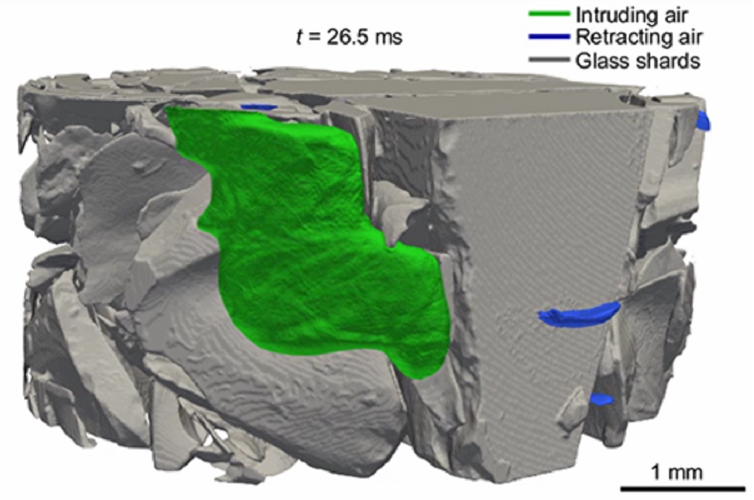
Towards the end of a Haines jump, a pore has been filled with encroaching air. This air is shown in green, while the surrounding material is grey. Illustration: NTNU
- You might also like: Glass fibres potential far beyond transmitting light
Important for medicine and CO2 storage
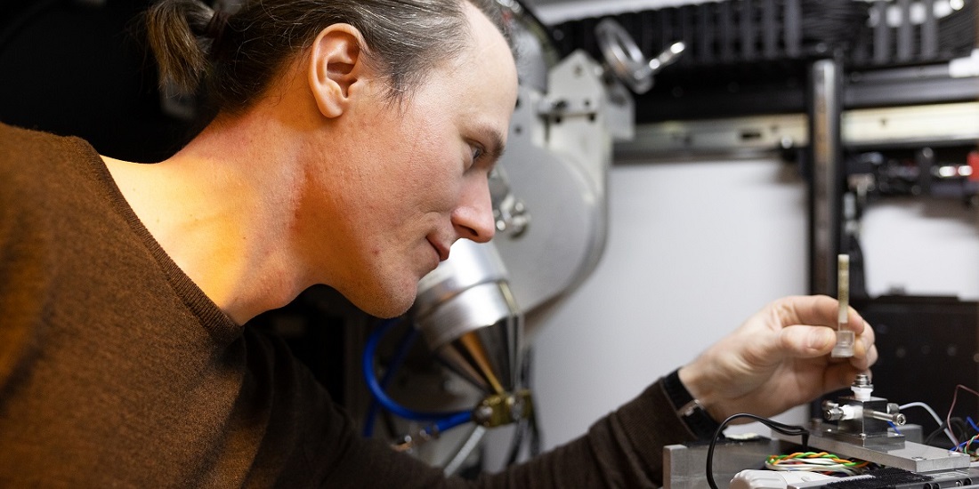
Kim Robert Tekseth is a doctoral student at NTNU. He is studying how X-ray microscopy can be used to study fluids in porous materials. Scientists around the world have been competing to make a slow motion 3D video of fluids in stone. The previous ‘world record’ was approximately one second per time step. Our research team has broken this record. We can now make these measurements around 1000 times faster! At 0.5 milliseconds per step, fluid flow can be studied in detail in 3D.
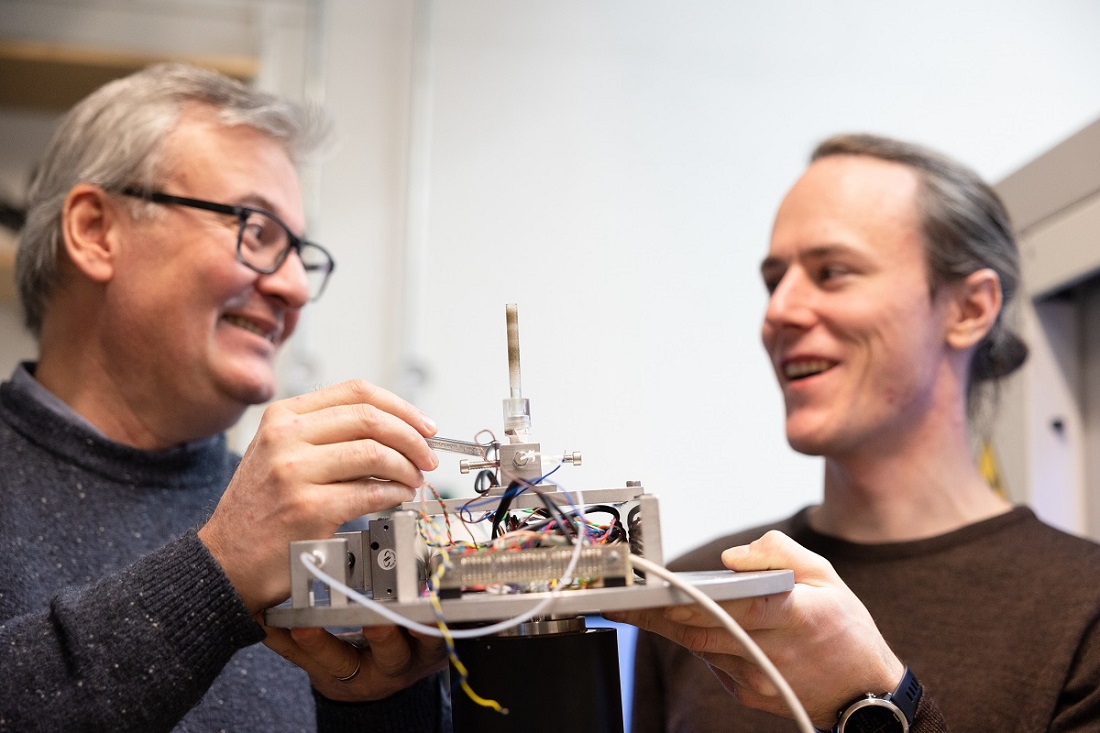
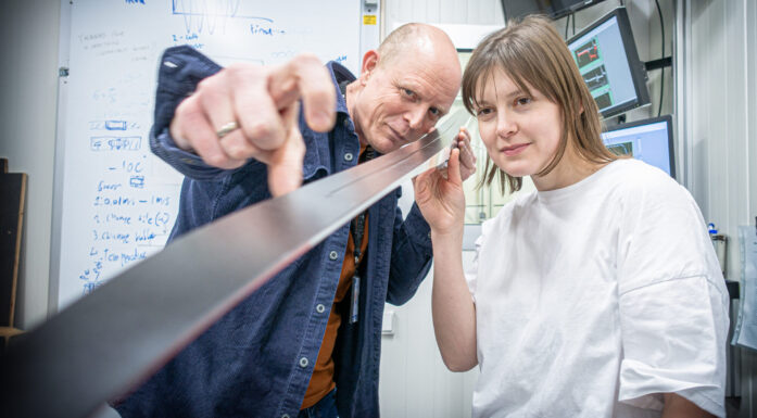
Professor Dag Werner Breiby and PhD candidate Kim Robert Tekseth with the test cell they used to study drainage. Photo: Per Henning, NTNU
Had to rethink entire process
Using regular CT, the sample must be rotated 180o to create each 3D-image. This limits the imaging rate, meaning we had to rethink the entire process. The solution was to make the flow through the porous material repeatable. We made a small sample of sintered glass. Water and air can be repeatedly driven back and forth inside the glass, while hundreds of thousands of X-rays are taken from different angles. The method can be illustrated by comparing it to the high jump in athletics.
Imagine that you are going to make a 3D film of a professional high jump. Multiple cameras can be used at the same time from different angles (but this is difficult to do with X-rays). The key is that each jump proceeds with near-identical technique each time. This enables you to record a series of jumps from different angles, and these recordings can then be compiled into a single 3D-film. This is also called 4D-CT (3D + time). The collaboration with the ESRF X-ray facility (synchrotron) in France played a crucial role.
This enabled us to measure that the liquid front moves during jumps by up to 200 mm/s, which is very much higher than the average flow rate. We also saw that when a pore suddenly filled during a jump, the fluid level was simultaneously affected in all the other pores of the sample. Our study is the first time this has been directly observed in 3D.
In the future, we will also be able to use our method in other rapid 3D processes. In addition to basic fluid studies, we will study catalysis and batteries. We have also used artificial intelligence to analyse the measurements faster and better.
The Research Council of Norway funded the work, and a recent research article has now been published in PNAS.
Reference: Kim Robert Tekseth, Fazel Mirzaei, Bratislav Lukic, Basab Chattopadhyay, and Dag Werner Breiby. Multiscale drainage dynamics with Haines jumps monitored by stroboscopic 4D X-ray microscopy. Proceedings of the National Academy of Sciences of the United States of America (PNAS). December 26, 2023, 121 (1) e2305890120 https://doi.org/10.1073/pnas.2305890120